The Neuronal Project
Our neuronal project also involves the use of microelectrode array to study the important electrophysiological changes that occur in the absence of presence of pharmacological interventions.
Past and present work includes the use of brain stem, cortex, and retinal ganglion cells. We often take advantage of the fact that neuronal cells tend to 'self-assemble' and they are moved around as the nerve fibres grow (growth cone). Some examples are shown below.
Past and present work includes the use of brain stem, cortex, and retinal ganglion cells. We often take advantage of the fact that neuronal cells tend to 'self-assemble' and they are moved around as the nerve fibres grow (growth cone). Some examples are shown below.
Brain cells will move away from a brain slice over time. |
Neuronal cell migration happens rapidly. |
Brain stem slice on MEA forming a diffused network.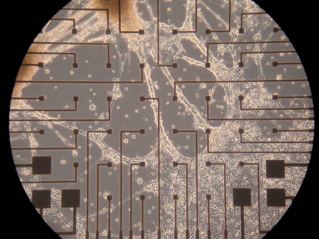
|
Migrating brain stem neurons on electrodes.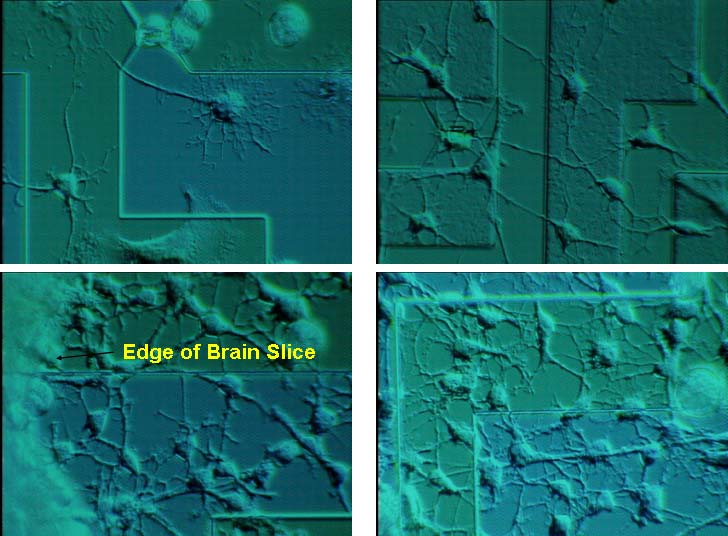
|
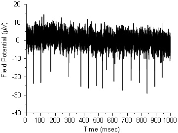
Spontaneously firing brain stem neurons.
|
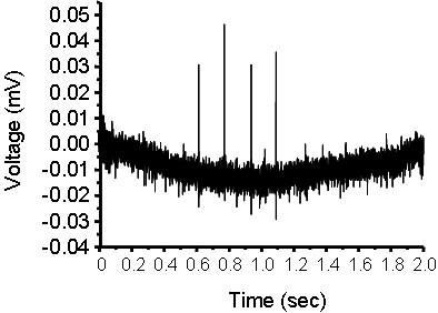
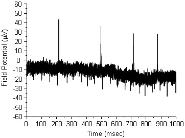
The firing rate of brain stem neurons increases upon the addition of rubidium chloride, which causes membrane depolarisation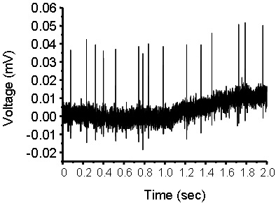
|
Neural Network
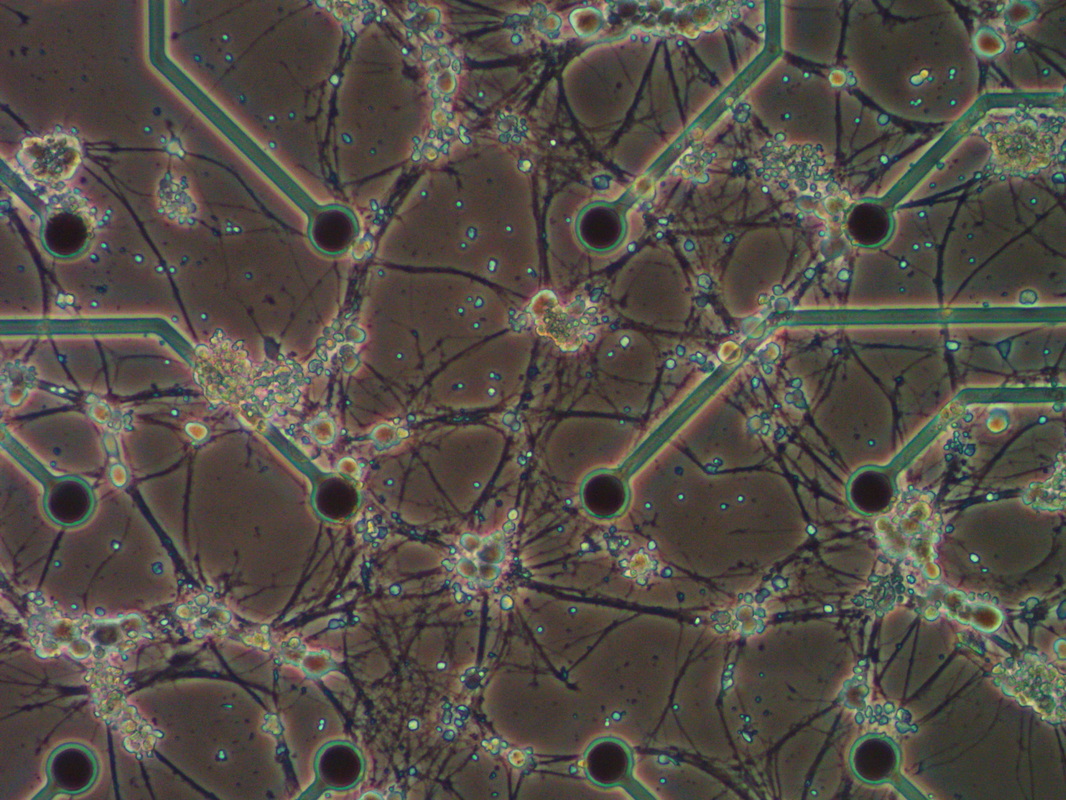
Apart from simply having either dissociated neurons or cells from explant cultures, we have been able to successfully form live neural networks.
3, 4 days old (Upper Left, Upper Right) and 8 days old (Lower Left, Lower Right) in culture. Migrated neurons from an initially 250mm thick brain slice.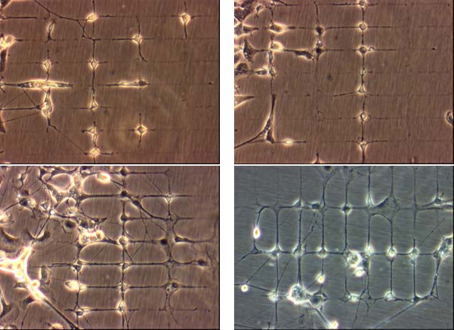
|
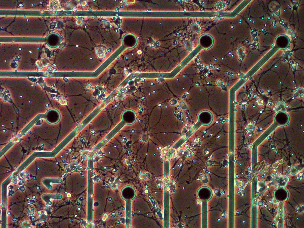
At higher magnification (Lower Left and Right). Neurons can be seen making direct connections with each other (9 days in culture), forming clear and well-defined connections.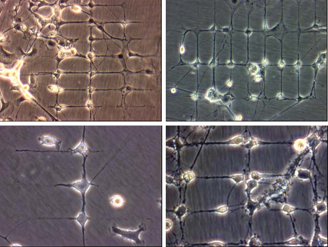
|
After 14 days in culture, It is possible that these formed nets can be directly placed onto microelectronic recording devices.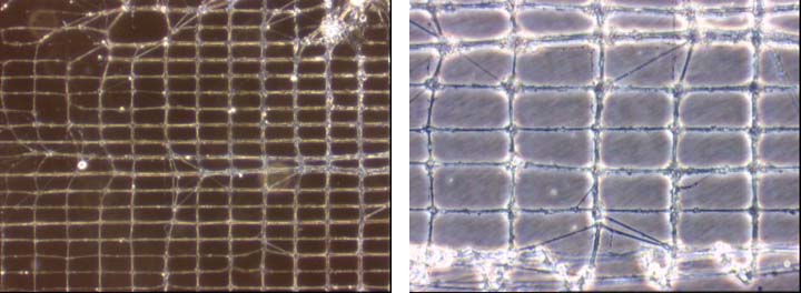
|
Polar tracers can be used to visualise cellular connections: Sulforhodamine, Cascade Blue, Lucifer Yellow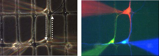
|
There are other neuronal cells that can be used, especially if they are spontaneously active. One such site is the retina, or more precisely, the retinal ganglion cells.
Retinal Ganglion Cells
We have begun a study on retinal ganglion cells (RGCs) in collaboration with colleagues at the Department of Ophthalmology and Visual Sciences (DOVS, CUHK). We will be studying the electrophysiology of RGCs in relation to ageing and other ocular ailments.
|
|
Depending on where the retinal ganglion cells are situation relative to the recording electrodes, different patterns of recording can be obtained.
|
|